
Age Spot & Bumps Treatment in Newport Beach, CA
Conveniently located to serve Newport Beach and Orange County
Personalized Non-Surgical Treatments for Age Spots, Skin Growths, and Sun Damage Offered in Orange County
Patients interested in age spots and bumps treatment in Newport Beach, CA, choose The Ocean Clinic Dermatology & Plastic Surgery. Board-certified dermatologist Dr. Amy Reinstadler and board-certified physician assistant Sarah Baca, PA-C, provide expert evaluation and treatment for the full range of age-related spots, growths, and textural changes that Orange County residents experience.

What Are Age Spots and Bumps?
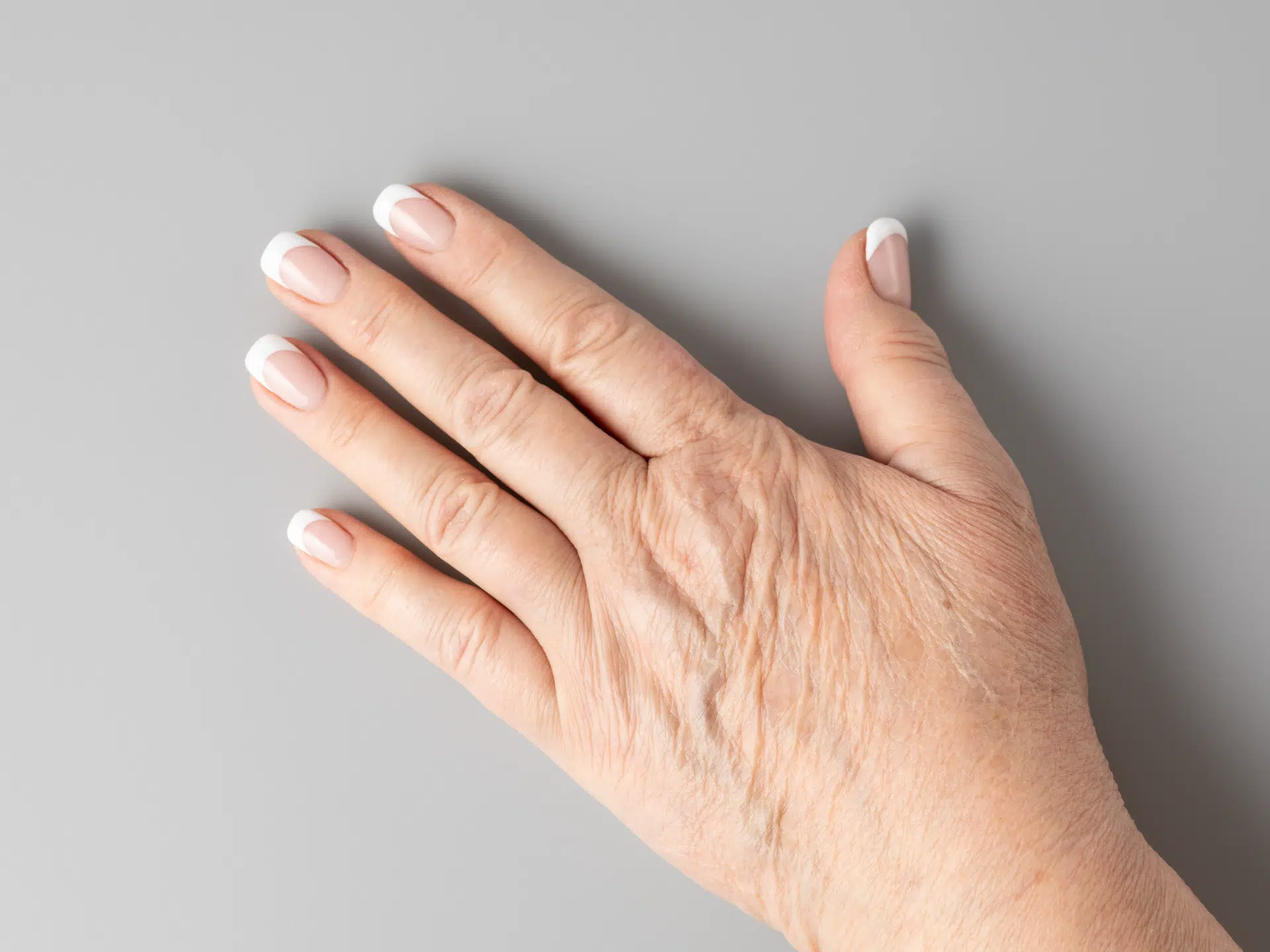
As the body matures, the skin’s appearance and characteristics change. Aging is accelerated in those areas exposed to sunlight, a process known as photoaging. Sun exposure, chronological aging, and even cigarette smoking all contribute to visible changes in the skin over time.
Types of Age Spots and Bumps
Signs of aging skin can be broken into three main categories: pigmentary (color) changes, blood vessel abnormalities, and growths/textural changes.
Pigmentary Changes
- Solar lentigines — Commonly known as age spots or liver spots, these are benign (non-cancerous) sun-induced lesions. They can be lightened with topical creams or treated in-office with lasers, liquid nitrogen, or light chemical peels.
- Guttate hypomelanosis — These lesions are “white freckles.” They’re small, flat, white spots most often found on the shins and forearms, though they may also appear on other sun-exposed areas. Sunscreen helps prevent new spots, and laser treatments may improve existing ones.
Blood Vessel Abnormalities
- Telangiectasias — Telangiectasias are widely open (dilated) blood vessels in the outer layer of the skin. When seen on the legs, they are often called spider veins. They appear as fine red, blue, or purple lines on the face or legs and may be treated with lasers or sclerotherapy.
- Cherry angiomas — Small red, purple, or nearly black vascular growths that tend to multiply after age 30. Cherry angiomas are harmless, but sometimes they can look similar to skin cancer. They can be removed with electrocautery or laser if desired.
- Solar (actinic) purpura — Solar purpura is a common condition in aging skin that appears as purple, red, or blue patches on the backs of the arms and hands. It results from sun-damaged connective tissue that can no longer adequately support small blood vessels. Daily sunscreen and topical tretinoin can help prevent further damage.
Growths/Textural Changes
- Solar elastosis — Solar elastosis is a skin condition that appears yellow and thickened as a result of sun damage. Treatment options include ablative and non-ablative lasers, dermal fillers, and topical tretinoin, along with sun protection and smoking cessation.
- Solar comedones — Whiteheads, blackheads, and occasionally even larger cystic lesions may develop on the face, particularly around the eyes, nose, chin, and ears, as a result of chronic sun exposure. In-office extractions and topical tretinoin are the primary treatments.
- Sebaceous hyperplasia — A condition in which sebaceous glands enlarge and protrude above the skin’s surface, forming small yellow bumps with a central indentation on the forehead, cheeks, or nose. Treatment options include surgical excision, electrocautery, cryotherapy, or laser therapy.
- Seborrheic keratoses — Seborrheic keratoses are common skin growths. They may look worrisome, but they are benign (not cancer). They have a waxy, stuck-on appearance and range from white to black in color, and are typically treated with liquid nitrogen or electrocautery and curettage.
- Skin tags — Skin tags are very common, soft, harmless lesions that appear to hang off the skin. They’re most often found in skin folds and can be removed for cosmetic reasons by cryotherapy, surgical excision, or electrosurgery.
Diagnosing Age Spots and Bumps
Diagnosis begins with a careful visual examination in our Newport Beach office of the affected areas, along with a review of your medical history, skin type, sun exposure history, and any treatments you may have already tried. In most cases, the appearance of the lesion is enough to reach a confident diagnosis. Occasionally, a biopsy or additional testing may be recommended to rule out more serious conditions, particularly when a growth has changed in size, color, or shape.
Treatment Options at Our Newport Beach Location
No two patients present with the same combination of concerns, which is why Amy Reinstadler, MD, and Sarah Baca, PA-C, take a highly individualized approach to every treatment plan. Depending on your diagnosis, care may involve topical therapies, prescription medications, or in-office procedures such as laser treatments, liquid nitrogen, electrocautery, or excision. Your provider will take the time to explain your options clearly and help you choose the approach that best fits your skin, lifestyle, and goals.
The Recovery Process
Recovery varies depending on the type and extent of treatment. Most in-office procedures allow patients to return to their normal routine that same day or shortly after. Some treated areas may need brief wound care or gentle handling while they heal, and follow-up visits might be scheduled to monitor progress or address additional lesions.
Benefits of Age Spot and Bump Treatment
- Clearer, more even-toned skin
- Reduced the appearance of sun damage and textural irregularities
- Early identification of potentially concerning growths
- Personalized care plans from experienced dermatology providers
- Minimized risk of lesion progression or spread
How Much Do Age Spot and Bump Treatments Cost?
Treatment price varies based on the types and number of lesions present, which procedures are recommended, and your insurance coverage. During your consultation, we’ll provide a detailed cost estimate and review any applicable insurance benefits.
Frequently Asked Questions
Can these conditions affect people with darker skin tones?
Yes, age spots, skin tags, seborrheic keratoses, and other growths affect people of all skin tones, though their appearance may differ. In darker skin, certain conditions can appear more deeply pigmented, and treatment requires careful selection to avoid post-inflammatory hyperpigmentation.
Schedule Your Appointment in Newport Beach, CA
Ready to address your age spots or bumps? The Ocean Clinic offers trusted dermatology care close to home in Orange County. Contact us today to schedule an appointment with Dr. Amy Reinstadler or Sarah Baca, PA-C, in our Newport Beach office.