
Mohs Micrographic Surgery Newport Beach
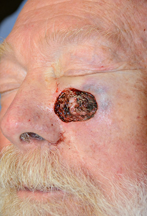
Mohs micrographic surgery is the excision of a cancer from the skin, followed by the detailed mapping and complete microscopic examination of the cancerous tissue and the margins surrounding it. If the margins are cancer-free, the surgery is ended. If not, more tissue is removed, and this procedure is repeated until the margins of the final tissue examined are clear of cancer.
Mohs surgery eliminates the guesswork in the removal of skin cancers and pinpoints the cancer’s location when it is invisible to the naked eye. Mohs surgery differs from other techniques since the microscopic examination of all excised tissues during the surgery eliminates the need to estimate how far out or deep the roots of the skin cancer go. This allows the Mohs surgeon to remove all of the cancer cells while sparing as much normal tissue as possible.
The cure rate of the Mohs technique is 99 percent for most skin cancers, considerably higher than that of other methods; it also provides the greatest chance of cure when other methods have failed.
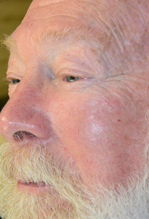
It is important to note that Mohs surgery is not appropriate for the treatment of all skin cancers. Mohs micrographic surgery typically is reserved for those skin cancers that have recurred following previous treatment or for cancers that are at high risk for recurrence. Mohs surgery also is indicated for cancers located in areas such as the nose, ears, eyelids, lips, hairline, hands, feet, and genitals, in which maximal preservation of healthy tissue is critical for cosmetic or functional purposes.
Mohs surgery can take a few hours to the better part of a day depending on how large the cancer is and how deep its “roots” go. Mohs surgery tends to be so time-consuming because a lot of the work is done behind the scenes while the patient waits. After the excision, the surgeon marks the tissue with colored ink to maintain orientation of the tissue when it is viewed under the microscope. Then a meticulously drawn map of the inked tissue is made; this will be used to document the location of any remaining cancer. The tissue is sliced into wafer-thin sections, which are placed on a glass slide. The slides are then stained to make any residual cancer visible. The physician examines the slides, indicating the exact location of any remaining cancer on the map. Finally, the doctor uses the map to pinpoint the exact location of the cancer on the patient and then excises any remaining cancerous tissue. This process is repeated until complete removal of the cancer is confirmed.
Dr. Amy Reinstadtler will see you in her office in Newport Beach.